As an eye doctor, it is easy to forget how foreign the anatomy of the eye can be for the layperson. We don’t mean to confuse you when we talk to you about your eye ball anatomy and your eye health, but sometimes we forget that we have been studying and living eyeballs for years and even decades and you haven’t had that same exposure.
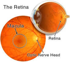
So let’s talk about the inside of the eye for a moment. Let’s talk about the retina. In the picture to the left anything that is orange is the retina. The photographs that you see are a true representation of what the retina looks like. You will also notice the optic nerve head that is round and somewhat whiter than the surrounding retina and you will notice that the macula has been circled. The macula is the central part of the retina, it is the most important part of the retina. The macula provides us with our detailed vision and our color vision. The rest of the retina provides us with peripheral vision, but is not able to give us detail about our world.
The retina and the macula both are very thin and delicate membranes. They are about the constancy of wet tissue paper. They are composed of millions of little tiny biological cameras we call rods and cones. The rods are good at motion detection and low light vision and the cones are better at detail and color discrimination. As you might guess, cones populate the macula, rods predominate in the peripheral retina.